
Three Common Hoof Problems in Dairy Cows:
What to Look for and How to Prevent Them
Key Takeaways
- Hoof lesions are the leading cause of lameness in dairy cows.
- The most common hoof lesions are digital dermatitis, sole ulcers, and white line disease.
- Each lesion develops differently and requires a specific prevention and treatment approach.
- Early detection and treatment improve recovery and help limit disease progression.
- Prevention focuses on hygiene, cow comfort, hoof trimming, and reducing stress on the hoof.
This fact sheet covers the three most common hoof lesions in dairy cows and explains why they matter and how to prevent them on your farm.
Why Talk About Hoof Lesions?
Lameness means a cow is not walking normally, and hoof problems are the most common cause. Lameness can also result from other conditions, such as trauma, arthritis, muscle or tendon injuries, and neurologic disease (Cramer et al., 2024). Most lame cows have an underlying hoof lesion, but not all cows with hoof lesions will appear visibly lame, especially when lesions are mild or in the early stages (van Huyssteen et al., 2020).
The three most common hoof lesions in North American dairy herds are digital dermatitis, sole ulcers, and white line disease (Cramer et al., 2008; DeFrain et al., 2013; Solano et al., 2016). Because these lesions are so common, they are an important place to focus prevention efforts.
Hoof lesions and lameness can have serious effects on cow health and herd performance. Lameness is associated with lower milk production (Puerto et al., 2021), poorer reproduction (Mellado et al., 2018), and a greater risk of culling (Ózsvári, 2017; Puerto et al., 2021). Preventing hoof lesions and treating them early can help reduce these losses.
Understanding the most common hoof lesions is the first step to spotting problems early and improving hoof health on your farm. For a broader overview of lameness and how to recognize it on your farm, see the USU Extension fact sheet “Lameness in Dairy Cattle: A Practical Overview.”
For a more technical overview of these hoof lesions, see USU Extension’s “Digital Dermatitis, Sole Ulcers, and White Line Disease in Dairy Cattle: Pathogenesis, Diagnosis, and Treatment.”
Digital Dermatitis
What is it?
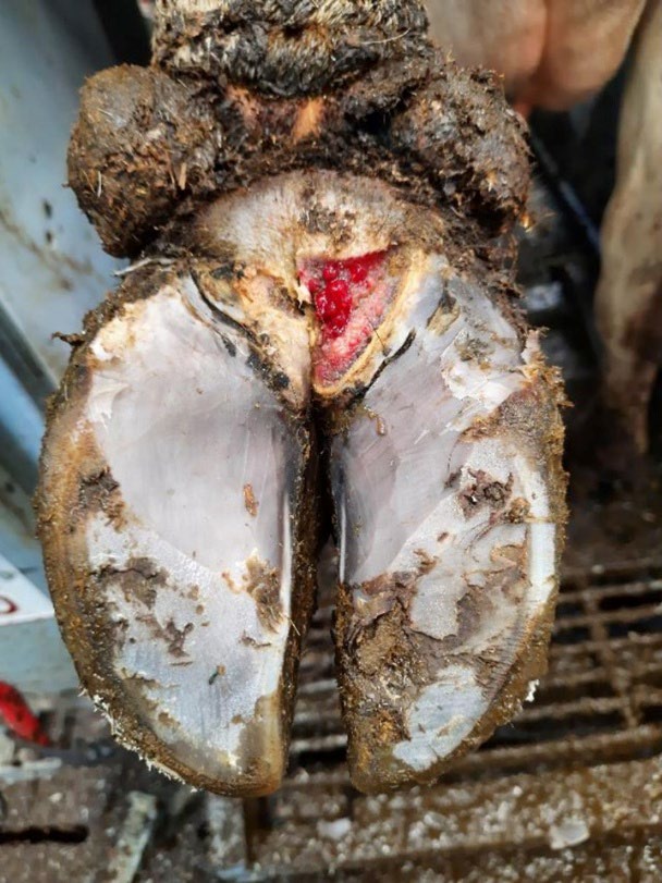
Digital dermatitis is an infectious skin lesion that most often occurs in the area between the heel bulbs of the foot (Figure 1; Read & Walker, 1998), but it can be found in other locations (Hernandez & Shearer, 2000).
It is commonly called foot warts or hairy heel warts, but digital dermatitis is the standard term (Orsel et al., 2018). Digital dermatitis was first described in 1974 (Cheli & Mortellaro, 1974) and is one of the most common causes of lameness in dairy herds.
Why does it happen?
Digital dermatitis develops when the skin on the foot is damaged, most often from constant exposure to moisture, manure, and abrasive conditions (Cramer & Solano, 2024). Once the skin is weakened, bacteria can enter and cause infection.
Treponema species are commonly found in these lesions and play a key role (Mamuad et al., 2020). As the infection spreads, it damages the skin and causes inflammation, leading to painful sores or thickened, overgrown tissue (Döpfer et al., 1997; Berry et al., 2010).
What does it look like?
Digital dermatitis lesions are often described using the M-stage system, which classifies lesions based on how active they are and how they appear (Figure 2; Döpfer et al., 1997; Berry et al., 2012).
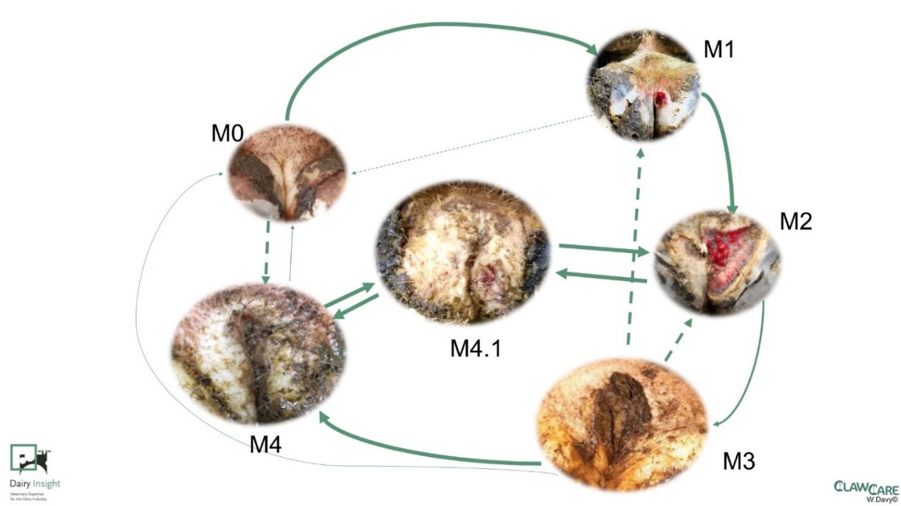
Sources: Döpfer et al., 1997; Berry et al., 2012
Early or active lesions are usually small, raw, and painful areas on the skin. These can develop into larger, open sores that are very sensitive and may cause lameness. As lesions begin to heal, they form scabs. Chronic lesions are typically thicker, more developed, and may be less painful, but they can still flare up and become active again. Understanding whether a lesion is active or chronic is important because it helps guide treatment and management decisions.
How can you prevent it?
Preventing digital dermatitis starts with keeping feet clean and dry. Reducing exposure to manure, moisture, and abrasive surfaces helps protect the skin and lowers the risk of infection (Cramer & Solano, 2024).
Footbaths are the most common prevention tool. They are typically filled with products such as copper sulfate or formalin and should be long enough to allow at least three steps per rear foot, usually 10 to 12 feet (Cook, 2017). The goal of a footbath is reducing the number of active, painful lesions, with frequency adjusted based on how common digital dermatitis is in the herd.
Biosecurity also plays an important role. Higher digital dermatitis rates have been linked to herds where animals were recently purchased and to unsanitized boots from employees or visitors (Oliveira et al., 2017). Screening incoming animals and ensuring proper boot sanitation can help reduce the risk of introducing and spreading the disease.
How is it treated?
Treatment focuses on active lesions that are painful and affect the cow. These lesions respond best to prompt, individual treatment (Solano et al., 2017). Less active or chronic lesions may be managed through herd-level control strategies, such as footbaths, rather than individual treatment (Bell & Vanhoudt, 2020).
Active digital dermatitis lesions are most often treated with topical products applied directly to the lesion. These include antibiotic products, such as tetracycline-based powders or pastes, as well as non-antibiotic options that often contain copper or other compounds. Because approved products and regulations vary, work with your herd veterinarian to choose the right treatment and follow withdrawal guidelines (Cramer & Solano, 2024).
Before treatment, clean the foot to remove manure while avoiding additional damage or bleeding. After applying treatment, a wrap may be used but is not required. If used, remove wraps within 24 hours to avoid creating additional problems (Cramer & Solano, 2024).
Sole Hemorrhage and Sole Ulcer
What are they?
Sole hemorrhages and sole ulcers are closely related hoof problems that develop in the same area of the foot, most often on the outside hoof of the rear feet (Shearer & Amstel, 2017). The main difference between them is severity. Sole hemorrhages appear as red, purple, or yellow discoloration in the sole and are often an early sign of damage. Sole ulcers are more severe and occur when normal hoof horn production is disrupted, leading to a defect in the sole that exposes the sensitive tissue underneath.
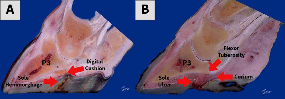
(A) Sole hemorrhage with labeled structures including the third phalanx (P3), digital cushion, and hoof horn defect.
(B) Sole ulcer with labeled structures including the third phalanx (P3), corium, and flexor tuberosity of the P3.
Why do they happen?
Sole hemorrhages and sole ulcers develop when too much pressure from the bone inside the hoof (third phalanx; P3) is placed on the horn-producing tissue, or corium (Figure 3, A and B; Shearer & Amstel, 2017). This pressure increases when hoof support is weakened, such as around calving when hormones make the hoof’s supporting structures more flexible (Tarlton et al., 2002), or when cows have less digital cushion to absorb impact (Newsome et al., 2017).
Early damage causes bleeding within the corium, which appears as discoloration in the sole. If pressure continues over time, normal horn production is disrupted, leading to a defect in the sole that allows the underlying corium to become exposed, resulting in a sole ulcer (Shearer & Amstel, 2017).
What do they look like?
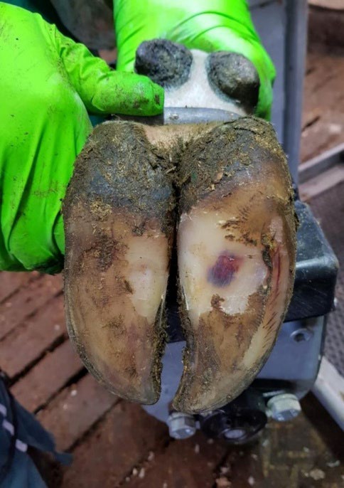
Sole hemorrhages appear as areas of discoloration in the sole, ranging from red and yellow to blue or purple (Figure 4; Cramer & Solano, 2024). These lesions often develop before a cow shows signs of lameness, so they are commonly found during routine hoof trimming rather than by watching how a cow walks.
How can you prevent them?
Preventing sole hemorrhages and sole ulcers focuses on reducing excess pressure on the horn-producing tissue (corium), especially from the P3 (Cramer & Solano, 2024).
One of the most important strategies is promoting adequate lying time. Cows should spend about 12 to 14 hours per day lying down, and time away from resting areas should be limited to no more than 3 to 4 hours per day (Cramer & Solano, 2024). More time standing increases pressure on the hoof and raises the risk of these lesions.
Maintaining body condition is also critical. Cows that lose too much condition, especially around calving, have less digital cushion to absorb pressure, increasing their risk of hoof damage (Cramer & Solano, 2024).
Proper hoof trimming is another key tool. Correct trimming helps balance weight across the hoof and reduces areas of high pressure (Cramer & Solano, 2024).
Many factors influence standing time and body condition, including stall comfort, stocking density, flooring, heat stress, transition cow management, and nutrition. Addressing these areas together is important for effective prevention (Cramer & Solano, 2024).
How are they treated?
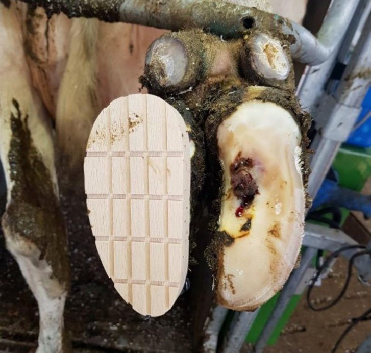
Treating sole hemorrhages depends on lesion severity and whether the cow is in pain (Cramer & Solano, 2024). Non-painful hemorrhages identified during routine trimming can often be managed by lowering the heel to reduce pressure beneath the P3. In contrast, hemorrhages that persist during trimming and cause a withdrawal response to hoof testers should be treated more aggressively by applying a hoof block to the unaffected hoof to reduce weight bearing on the affected area (Figure 5).
Sole ulcers are treated by carefully removing loose or undermined hoof horn around the exposed corium to prevent debris buildup and allow healing (Cramer & Solano, 2024). A hoof block should then be applied to the unaffected hoof to offload weight from the affected area and support horn regrowth.
Cows fitted with hoof blocks should be re-evaluated within 3 to 6 weeks to assess healing and adjust or remove the block as needed. Also consider using non-steroidal anti-inflammatory drugs (NSAIDs). Inflammation associated with these lesions has been linked to bony growths of the P3 (Newsome et al., 2016) and deterioration of the digital cushion (Wilson et al., 2021), which may increase pressure on the corium and raise the risk of recurrence.
White Line Disease
What is it?
White line disease includes a range of problems that affect the white line, the area where the hoof wall and sole meet. These problems can vary in severity and include cracks, separations, and abscesses (Cramer & Solano, 2024).
- Fissures are small cracks in the white line.
- Separations occur when the hoof wall and sole begin to pull apart.
- Abscesses develop when bacteria enter through a crack or separation, leading to infection and buildup of pus.
Why does this happen?
White line disease develops when the white line becomes weakened and more prone to damage. The white line is naturally softer than the surrounding hoof, which makes it more vulnerable to stress (Shearer et al., 2015).
Similar to sole ulcers, problems begin when hoof support is weakened. This allows the P3 to place more pressure on the corium, which can interfere with normal horn formation (Shearer & van Amstel, 2017). As the white line weakens, cracks or separations can form. These openings allow dirt, manure, and bacteria to enter, leading to inflammation and, in more severe cases, abscess formation (Cramer & Solano, 2024).
What does it look like?
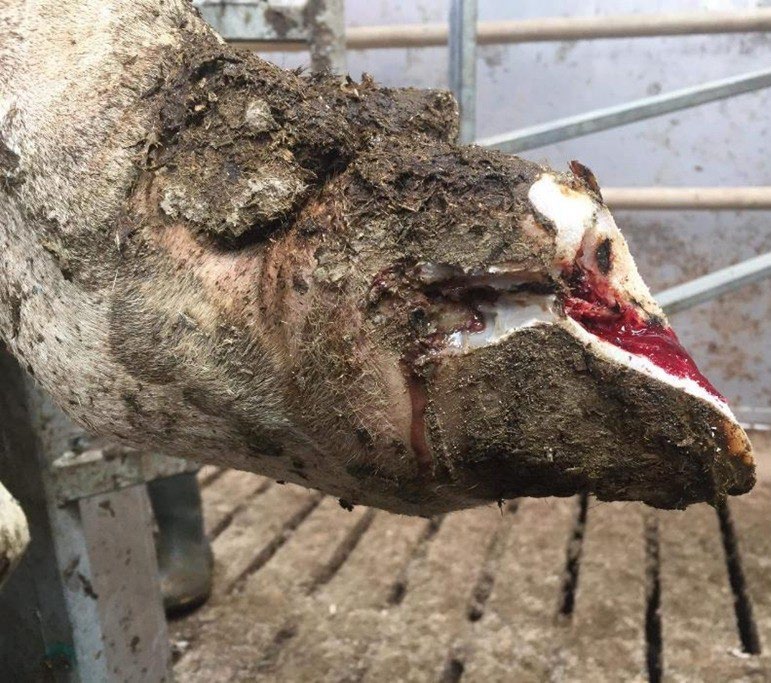
White line disease can appear as hemorrhage, separation, or abscess formation and may or may not cause visible lameness (Figure 6; Cramer & Solano, 2024). Early lesions may show up as small separations in the white line at the sole. Over time, these can trap dirt and bacteria and develop into abscesses. In more advanced cases, abscesses may create draining tracts that move upward toward the coronary band or heel.
These lesions are most commonly found along the outer edge of the white line in weight-bearing areas of the hoof, although they can occur anywhere along the white line.
How can you prevent it?
Preventing white line disease focuses on reducing stress on the white line, especially from excessive force or twisting of the hoof (Cramer & Solano, 2024).
- Flooring is one of the most important factors. Surfaces should provide good traction without being overly abrasive or slippery. Rubber flooring and properly designed grooves can help reduce stress on the hoof.
- Facility design and cow movement also matter. Cows should be able to move calmly and at their own pace. Sharp turns, overcrowding, and rushed handling increase pressure and twisting forces on the hoof.
- Consistent and well-timed hoof trimming is another key tool. Proper trimming helps balance weight across the hoof and reduces stress on the white line.
How is it treated?
Treating white line disease depends on lesion severity and is similar to that of sole ulcers (Cramer & Solano, 2024). Treat painful lesions by removing loose or damaged hoof horn around the lesion, including affected wall horn. The heel may be lowered, and a hoof block applied to the unaffected hoof to offload weight from the affected area (Figure 5). This allows the corium to heal and supports normal horn regrowth.
Cows fitted with hoof blocks should be re-evaluated within 3 to 6 weeks to assess healing and adjust or remove the block as needed.
Using non-steroidal anti-inflammatory drugs (NSAIDS) should also be considered. Inflammation associated with these lesions can lead to changes in the P3 and the digital cushion that may increase pressure on the corium and raise the risk of recurrence (Newsome et al., 2016; Wilson et al., 2021). Additional information on this process is provided in the Sole Ulcer section.
Summary
Hoof lesions are the leading cause of lameness in dairy cows, with digital dermatitis, sole ulcers, and white line disease being the most common. Although these conditions may appear similar, they develop for different reasons and require different approaches to prevention and treatment. Understanding how each lesion forms improves early detection and management decisions on the farm. Focusing on hygiene, cow comfort, proper hoof trimming, and minimizing stress on the hoof can reduce the risk of these lesions and improve overall cow health and performance.
For a more detailed, technical explanation of these lesions, see the companion USU Extension fact sheet “Digital Dermatitis, Sole Ulcers, and White Line Disease in Dairy Cattle: Pathogenesis, Diagnosis, and Treatment.”
Acknowledgments
All figure images were used with the permission of William Davy. Dennis Hinkamp, USU Extension, provided the first-page photo.
The authors of this content used ChatGPT to improve writing clarity and flow. The tool was not used to identify sources, generate ideas, or interpret findings. Authors reviewed and edited the content provided by the AI tool, and they take full responsibility for the content.
Continued Reading and Works Cited
Berry, S. L., Read, D. H., Famula, T. R., Mongini, A., & Döpfer, D. (2012). Long-term observations on the dynamics of bovine digital dermatitis lesions on a California dairy after topical treatment with lincomycin HCl. The Veterinary Journal, 193, 654–658. https://doi.org/10.1016/j.tvjl.2012.06.048.
Berry, S. L., Read, D. H., Walker, R. L., & Famula, T. R. (2010). Clinical, histologic, and bacteriologic findings in dairy cows with digital dermatitis (footwarts) one month after topical treatment with lincomycin hydrochloride or oxytetracycline hydrochloride. Journal of American Veterinary Medical Association, 237, 555–560. https://doi.org/10.2460/javma.237.5.555.
Cheli, R., & Mortellaro, C. (1974). Lesions of the digital cushion in cattle. In Proceedings of the 8th International Conference on Diseases of Cattle (pp. 208–213).
Cook, N. B. (2017). A review of the design and management of footbaths for dairy cattle. Veterinary Clinics: Food Animal Practice, 33, 195–225. https://doi.org/10.1016/j.cvfa.2017.02.004.
Cramer, G., Lissemore, K. D., Guard, C. L., Leslie, K. E., & Kelton, D. F. (2008). Herd- and cow-level prevalence of foot lesions in Ontario dairy cattle. Journal of Dairy Science, 91, 3888–3895. https://doi.org/10.3168/jds.2008-1135.
Cramer, G., & Solano, L. (2024). Lameness originating in the hoof in cattle. In Merck veterinary manual. Accessed January 13, 2025, from https://www.merckvetmanual.com/musculoskeletal-system/lameness-in-cattle/lameness-originating-in-the-hoof-in-cattle.
Cramer, G., Solano, L., & Abuelo, A. (2024). Overview of lameness in cattle - musculoskeletal system. In Merck veterinary manual. Accessed February 10, 2026, from https://www.merckvetmanual.com/musculoskeletal-system/lameness-in-cattle/overview-of-lameness-in-cattle.
DeFrain, J. M., Socha, M. T., & Tomlinson D. J. (2013). Analysis of foot health records from 17 confinement dairies. Journal of Dairy Science, 96, 7329–7339. https://doi.org/10.3168/jds.2012-6017.
Döpfer, D., ter Huurne, A. A. H. M., Cornelisse, J. L., van Asten, A. J. A M., Koopmans, A., Meijer, F. A., Schukken, Y. H., Szakáll, I., Klee, W., & Bosma, R. B. (1997). Histological and bacteriological evaluation of digital dermatitis in cattle, with special reference to spirochaetes and Campylobacter faecalis. Veterinary Record, 140, 620–623. https://doi.org/10.1136/vr.140.24.620.
Hernandez, J., & Shearer, J. K. (2000). Efficacy of oxytetracycline for treatment of papillomatous digital dermatitis lesions on various anatomic locations in dairy cows. Journal of American Veterinary Medical Association, 216, 1288–1290. https://doi.org/10.2460/javma.2000.216.1288.
Mamuad, L. L., Seo, B. J., Faruk, Md. S. A., Espiritu, H. M., Jin, S. J., Kim, W.-I., Lee, S.-S., & Cho, Y.-I. (2020). Treponema spp., the dominant pathogen in the lesion of bovine digital dermatitis and its characterization in dairy cattle. Veterinary Microbiology, 245, 108696. https://doi.org/10.1016/j.vetmic.2020.108696.
Mellado, M., Saavedra, E., Gaytán, L., Veliz, F. G., Macías-Cruz, U., Avendaño-Reyes, L., & García, E. (2018). The effect of lameness-causing lesions on milk yield and fertility of primiparous Holstein cows in a hot environment. Livestock Science, 217, 8–14. https://doi.org/10.1016/j.livsci.2018.09.008.
Newsome, R., Green, M. J., Bell, N. J., Chagunda, M. G. G., Mason, C. S., Rutland, C. S., Sturrock, C. J., Whay, H. R., & Huxley, J. N. (2016). Linking bone development on the caudal aspect of the distal phalanx with lameness during life. Journal of Dairy Science, 99, 4512–4525. https://doi.org/10.3168/jds.2015-10202.
Newsome, R. F., Green, M. J., Bell, N. J., Bollard, N. J., Mason, C. S., Whay, H. R., & Huxley, J. N. (2017). A prospective cohort study of digital cushion and corium thickness. Part 1: Associations with body condition, lesion incidence, and proximity to calving. Journal of Dairy Science, 100, 4745–4758. https://doi.org/10.3168/jds.2016-12012.
Oliveira, V. H. S., Sørensen, J. T., & Thomsen, P. T. (2017). Associations between biosecurity practices and bovine digital dermatitis in Danish dairy herds. Journal of Dairy Science, 100, 8398–8408. https://doi.org/10.3168/jds.2017-12815.
Orsel, K., Plummer, P., Shearer, J., De Buck, J., Carter, S. D., Guatteo, R., & Barkema, H. W. (2018). Missing pieces of the puzzle to effectively control digital dermatitis. Transboundary and Emerging Diseases, 65, 186–198. https://doi.org/10.1111/tbed.12729.
Ózsvári, L. (2017). Economic cost of lameness in dairy cattle herds. Journal of Dairy, Veterinary & Animal Research, 6. https://doi.org/10.15406/jdvar.2017.06.00176.
Puerto, M. A., Shepley, E., Cue, R. I., Warner, D., Dubuc, J., & Vasseur, E. (2021). The hidden cost of disease: II. Impact of the first incidence of lameness on production and economic indicators of primiparous dairy cows. Journal of Dairy Science, 104, 7944–7955. https://doi.org/10.3168/jds.2020-19585.
Read, D. H., & Walker, R. L. (1998). Papillomatous digital dermatitis (footwarts) in California dairy cattle: clinical and gross pathologic findings. Journal of Veterinary Diagnostic Investigation, 10, 67–76. https://doi.org/10.1177/104063879801000112.
Shearer, J. K., & van Amstel, S. R. (2017). Pathogenesis and Treatment of Sole Ulcers and White Line Disease. Veterinary Clinics: Food Animal Practice, 33, 283–300. https://doi.org/10.1016/j.cvfa.2017.03.001.
Shearer, J. K., Plummer, P. J., & Schleining J. A. (2015). Perspectives on the treatment of claw lesions in cattle. Veterinary Medicine: Research and Reports, 6, 273–292. https://doi.org/10.2147/VMRR.S62071.
Solano, L., Barkema, H. W., Mason, S., Pajor, E. A., LeBlanc, S. J., & Orsel, K. (2016). Prevalence and distribution of foot lesions in dairy cattle in Alberta, Canada. Journal of Dairy Science, 99, 6828–6841. https://doi.org/10.3168/jds.2016-10941.
Tarlton, J. F., Holah, D. E., Evans, K. M., Jones, S., Pearson, G. R., & Webster, A. J. F. (2002). Biomechanical and histopathological changes in the support structures of bovine hooves around the time of first calving. The Veterinary Journal, 163, 196–204. https://doi.org/10.1053/tvjl.2001.0651.
van Huyssteen, M., Barkema, H. W., Mason, S., & Orsel. K. (2020). Association between lameness risk assessment and lameness and foot lesion prevalence on dairy farms in Alberta, Canada. Journal of Dairy Science, 103, 11750–11761. https://doi.org/10.3168/jds.2019-17819.
Wilson, J. P., Randall, L. V., Green, M. J., Rutland, C. S., Bradley, C. R., Ferguson, H. J., Bagnall, A., & Huxley, J. N. (2021). A history of lameness and low body condition score is associated with reduced digital cushion volume, measured by magnetic resonance imaging, in dairy cattle. Journal of Dairy Science, 104, 7026–7038. https://doi.org/10.3168/jds.2020-19843.

July 2026
Utah State University Extension
Peer-reviewed fact sheet
Authors
Drew Swartz, Kalen Taylor, Jacob Hadfield, and Justin Clawson

Drew Swartz
Extension Assistant Professor | Dairy Cattle Specialist
Kalen Taylor
Extension Associate Professor with Tenure | Agriculture | Millard County Director
Agriculture and Natural Resources

Jacob Hadfield
Extension Associate Professor | Agriculture and Natural Resources | Utah and Juab Counties | Juab County Director
Agriculture and Natural Resources

Justin Clawson
Extension Assistant Professor | Agriculture & Natural Resources | Cache County
Agriculture and Natural Resources