Equine Piroplasmosis
Introduction
Equine Piroplasmosis (EP) is a tick-borne disease also referred to as babesiosis or theileriosis. This disease can affect horses, donkeys, mules, and zebras. Theileria equi and Babesia caballi are the causative parasitic agents of this disease which is spread by tick bites or through mechanical transmission by improperly disinfected instruments.1 Considered endemic (native to an area) in many tropical regions including Africa, Europe, the Middle East, Asia, Central and South America, EP is a foreign animal disease in the United States, and is not endemic here. Approxi-mately 14 species of ticks can be vectors for the organisms that cause this disease. Potential tick vectors for T. equi and B. caballi exist in the U.S.
Transmission
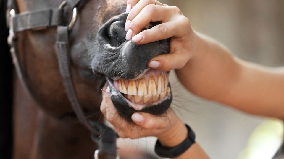
Equine Piroplasmosis occurs naturally when ticks ingest blood from an infected horse and then bite an uninfected horse, thereby spreading the parasite through blood contact. The disease replicates in the tick, making spread of the disease to multiple horses likely. Currently, there is only one known area in the U.S., located in southeast Texas, where tick transmission of EP agents has been shown to occur.1 Equine Piroplasmosis is also spread iatrogenically by humans using skin penetrating instruments without properly sanitizing these instruments or disposing of them between horses. These instruments include needles, syringes, dental equipment, tattoo equipment, and surgical tools.2 The majority of recent outbreaks in the United States have been attributed to iatrogenic transmission rather than the natural transmission via ticks.
Clinical Disease and Diagnosis
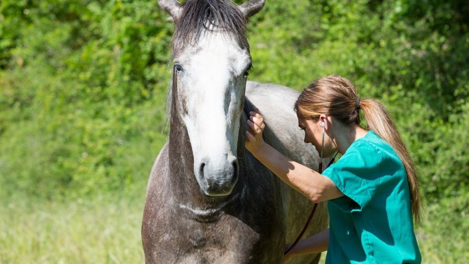
The blood borne parasites T. equi and B. caballi attach to the red blood cells and cause their destruction. After a horse is infected it can take from 7 to 30 days to show signs of the disease. Cases of EP may be unapparent, mild or acute. Mild cases can appear as loss of appetite or weakness, while more acute severe signs include fever, anemia, weight loss, jaundice, fluid accumulation in the abdomen, decreased performance, edema in the lower limbs, and even death. Because EP is not endemic in the U.S., our horses are naïve and highly susceptible to the acute forms of the disease.1 Mild forms of the disease may go completely unnoticed by horse owners. After the initial stages of the disease, EP may become chronic. Horses can carry the parasites for long periods of time with no clinical signs. This leads to a horse becoming a carrier that can spread the disease to other horses by mechanical or natural routes.
Because the clinical signs of EP are similar to other diseases, it can be challenging to diagnose without appropriate testing. The accepted method to diagnose EP is to test the serum or blood for antibodies to T. equi and B. caballi. Testing can take several days and may need to be sent to a national laboratory for confirmation testing. Due to the foreign animal disease designation in the United States, positive samples require notification of state and federal regulatory officials. Additionally, international notice to the World Organization for Animal Health (OIE) is conducted. The OIE is the international organizations that establishes standards for the safe international trade of animals.2
Today the United States enjoys the status of being free of EP from a travel and export perspective. The importance of protecting this designation cannot be understated when considering the negative economic impact the U.S. would incur if travel and trade were restricted as a result of allowing this disease to become endemic in the United States.
Prevention and Treatment
Current options for treating a horse testing positive for EP are limited. These options include life-long quarantine overseen by federal regulatory officials, euthanasia, and treatment. Treatment of EP is only permissible if the horse is enrolled in a USDA supervised research program. The goal of treatment is to completely clear the horse of infection. Although identification of a treatment protocol that can provide permanent clearance of EP from an infected animal would be ideal, it is not an outcome that can be guaranteed. The positive antibody status of the horse remains for many months and, in some cases, up to 2 years after successful treatment and the horse must remain quarantined during this time.
Prevention is vital to stop the spread of EP. In areas where the host tick is known to live, efforts to control ticks and keep them off horses have proven important. These ticks are one host ticks. They spend their life cycle on the same host as much as possible. In the United States, recent EP outbreaks have occurred in areas where the host tick is not known to live. These outbreaks have been isolated and caused by reusing needles and syringes between horses. The American Association of Equine Practitioners gives these guidelines:1
ALWAYS:
- Use a new sterile needle and syringe for injections, whether into a vein or muscle or beneath the skin.
- Carefully clean and disinfect equine dental, tattoo and surgical equipment between horses.
- Have any horse that will serve as a blood donor tested for EP.
- Contact your veterinarian if your horse is sick and has signs of fever, reduced feed intake or lethargy.
- Check with your State Animal Health Official if you need more specifics about EP.
Conclusion
Equine Piroplasmosis can be a devastating disease to horse owners and the horse industry. Understanding this disease and how it is spread will help us keep horses safe. With current outbreaks being limited to the race horse industry, the Utah Racing Commission has implemented a new commission order effective September 9, 2016, stating “no horse shall enter a sanctioned race track in Utah without first providing a negative Theileria equi cELISA test administered within the last twelve (12) months.” With these measures implemented in Utah and surrounding states, hopefully the current and potential threat EP poses to horses can be prevented.
References
- 1American Association of Equine Practitioners: Equine Piroplasmosis. Retrieved from http://www.aaep.org/info/horse-health?publication=758
- 2USDA APHIS 2009, Fact Sheet - Equine Piroplasmosis. Retrieved from https://www.aphis.usda.gov/publications/animal_health/content/printable_version/fs_equipir.pdf
Published October 2016
Utah State University Extension
Peer-reviewed fact sheet
Authors
Karl Hoopes, DVM USU Equine Extension Specialist; Barry Pittman, DVM Utah State Veterinarian

Karl Hoopes
Equine Specialist
ADVS Dept
Related Research
Aging Horses by Their Teeth
Being able to pinpoint age within a fairly narrow range can be of use to owners of unregistered horses or horses whose age is unknown for any reason. Many health and nutrition management decisions are directly related to age and dental wear, making it eve

Biosecurity at Equine Events: Event Committee Guidelines
Equine events have the potential for the perfect storm of equine disease spread. Horse’s immune systems are stressed by travel and competition, horses are comingled in close quarters, and various bacteria and viruses are brought from different locations.

Bitting the Horse: Snaffles
As horses are introduced to carrying a saddle and rider, they are also introduced to carrying a bit. The horse’s teeth should be evaluated prior to bitting. Points and hooks should be filed and wolf teeth should be removed. Wolf teeth are small, shallowro
Buying Your First Horse
Buying a horse can be quite a task for the first time horse buyer. It is very beneficial for the buyer to have some horse experience prior to ownership, perhaps by working at a stable or by taking riding lessons. Prior experience with horse management and
Caring for Horses in Cold Weather
During dark winter months, we often start to worry about our horses being outside in the cold. How do they stay warm? Horses adapt very well to colder weather. During the fall months, as temperatures cool gradually, horses begin to add additional fat and
Daily Horse Observations for Horse Owners
When responsible for a horse, an owner must be very observant. Knowing what is normal and what is not for each individual horse can alert the owner or caretaker to a problem before it becomes a big issue. Owners that have a routine, which can be followed